Who Invented the CT Scan?
Written by Manya Pandey, a first-year undergraduate student
CT scan, short for Computerised Tomography, is an imaging technique used to capture images within your body
Written by Manya Pandey, a first-year undergraduate student
Have you wondered – who invented the CT Scan?
First, let’s see what it is. A CT scan, short for Computerised Tomography, is an imaging technique used to capture images within your body. It uses X-rays to form 3-D images of objects.
It is widely used to scan and monitor diseases. Here’s everything about it…
How Do CT Scans Work?
A CT scanner is a tunnel-like machine that has a bowl-shaped apparatus attached to emit X-rays, called an X-ray emitter. A patient lies on a conveyor belt that slides them into the open-ended tunnel.
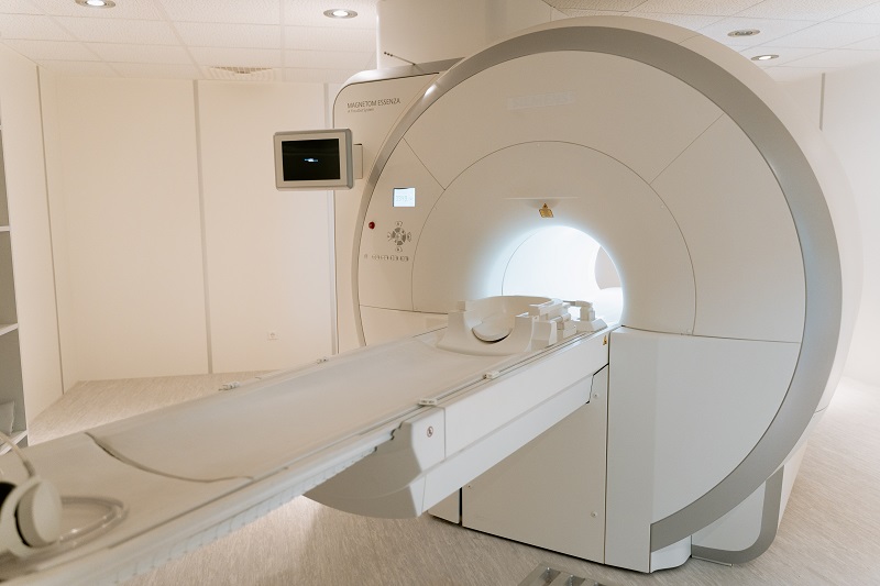
Inside the machine(tunnel), the X-ray emitter rotates in circles around the patient, taking up to 4-300 scans, sometimes up to 600
How is a 3-D image produced?
So, imagine stacking 300 square pages of the same size on top of each other. Shouldn’t you get a 3-dimensional solid box? Consider your textbooks. Individual pages inside them are 2-D, but stacked together, they’re a 3-D shape commonly known as a cuboid.
CT scans work on the same principle – more than hundreds of images of the same body part are taken quickly to create grey-and-white images. Computer software puts them together to form 3-D organs and diagnose the extent of the damage.
Say you broke a bone. Multiple scans of the injured area, placed on top of each other, will help the doctor determine how deep the fracture is!
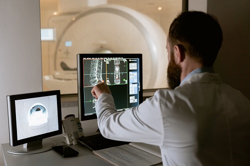
Different body parts absorb different amounts of radiation, so the images are created in shades of grey and white. This colour gradient also provides clarity to the doctors. For instance, bones appear brighter because they absorb less radiation.
What can CT scans detect?
- Broken bones
- Cancers
- Blood clots(when blood clumps up in the body)
- Signs of heart disease
- Internal bleeding (the bleeding we can’t see on the outside)
Your scan might use a dye
Special imaging is used to detect anomalies that are otherwise undetectable by a conventional CT scan. This is called CT perfusion imaging. In this technique, your scan highlights certain features using a contrast agent/ dye.
This type of CT may be used on the heart and brain. It is better for stroke diagnosis than other CT types.
The healthcare provider will either have you drink a special liquid containing the contrast agent or give you an injection, depending on the requirement. The contrast agent is cleared from your body through urine within 24 hours.
Does it pain?
No, the scanning procedure is completely painless. Moreover, it is completely automated, so all you have to do is lie on the conveyor belt and let it take you inside, you might find your head on the other side of the machine.
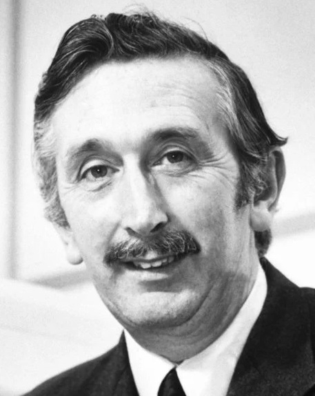
Who invented CT scans?
The person who developed the CT technique was an English electrical engineer named Godfrey Hounsfield. In 1979, he won a Nobel Prize in Physiology or Medicine along with Dr. Allan MacLeod Cormack for developing the diagnostic technique of X-ray computed tomography (CT).
However, here’s the thing..
Mathematically the idea was conceived in 1917 by William H. Oldendorf, an American neurologist, physician, and researcher who developed the technique of computed tomography.
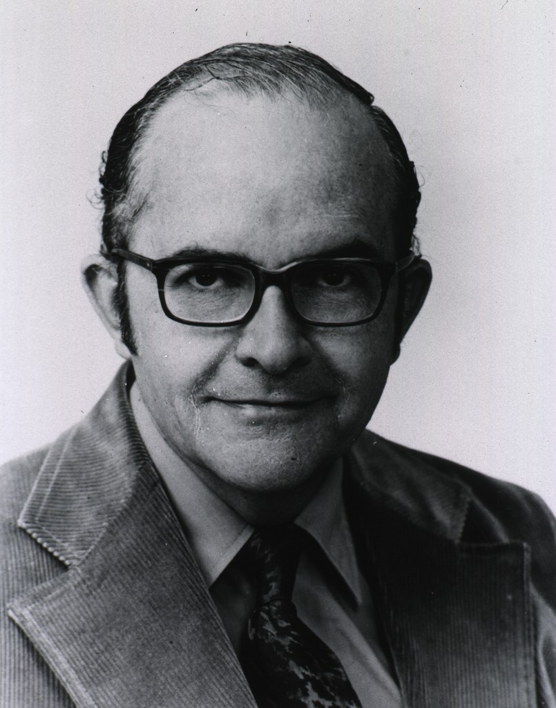
CT scans are based on his mathematical theory of the ‘Radon transform’ which he patented in 1963, as a “radiant energy apparatus for investigating selected areas of interior objects obscured by dense material”
How it began – an interesting backstory
The first commercially available CT scanner was launched by EMI Laboratories in 1972 as the “EMI brain scanner”.
According to the inventor Godfrey Hounsfield, the idea to build a CT scanner began on a vacation as his simple wish to reconstruct a 3D picture of a box. He intended to achieve this by re-imagining the object as a stacked series of 2-D slices.
This idea was taken further by EMI research. The project was funded and after a long journey of several decades, it transformed into what we know today as CT scanners.
The first clinical CT scanners were installed between 1974 and 1976. The original systems were dedicated to imaging the head only.
But added research and time over time led to the development of full-body scanners around 1976. CT was widely available by 1980. There are about 30,000 CT scanners installed worldwide. Radiographers or radiology technologists are investigating different fields using this technology of the 70s.
Other uses
Medical
Lately, CT scan has been used majorly in radiology(a medical discipline that uses scanned images to find and treat diseases), preventive medicine and screening of diseases. CT colonography has been proven effective for diagnosis in people with a high risk of colon cancer and heart disease.
Archaeological
X-ray CT and micro-CT scans are also used to conserve and preserve objects of historical importance. Using CT scans, conservators and researchers can determine the material composition and structure of the research object, without any additional harm.
Preservatory
In fossil research (fossils are preserved remains of ancient plants and animals in stone or amber), direct research and observation can be damaging and can degrade the sample. So CT is used to develop the internal structure of such fragile fossils before using harder tools to explore more.
CT scanning is deployed in the conservation of national and international ancient relics and museum artifacts.
Engineering
CT scanning is utilized in the industry for internal inspection of components without spitting them apart. It is employed in processes like flaw detection and failure analysis.
Side effects of CT scans
The technology has improved in terms of speed and accessibility. Faster scanning helps to eliminate effects from patient motion such as breathing, heartbeat, etc, but there’s still a long way to go in terms of safety.
The amount of radiation used for scans is not considered safe by many professionals around the world. Radiation causes several health problems including skin cancer.
- Not safe for pregnant women
- May damage your DNA
- The dye might cause an allergic reaction
Did you know…
- The first-ever human patient to benefit from the brain scanner was a woman believed to be suffering from a brain tumour.
- William Roentgen, the discoverer of X- rays, did not patent it.

Better Your Child’s G.K. In 3 Minutes – Get This Free Newsletter
Get fun facts, simple and easy news, quizzes, and lots of other interesting things to read in your mailbox – for free! It’s what we call GK-on-the-go!
I Kid You Not now has a large readership across India and also parts of the world. If you want to write for us, you can submit your story here. You can also apply to become a news anchor. Apply here


Comments